A new way to see disease

Learn about our end-to-end platform for mapping the molecular geography of tissue biopsies.
A biopsy can be a critical step in understanding the health status of a person with suspicious symptoms—from a lump under the skin, to spots on a chest x-ray—and is often required to determine the diagnosis or severity of many diseases. Essentially a collection of human tissue, each biopsy is rich with information, and yet pathologists are limited by today's technology to examine only a narrow band of those details, while many important pathological characteristics remain difficult to resolve. Because biopsies are very common procedures in healthcare, the impact of expanding pathologists' capacity to discern biologically-relevant features could translate into a much more information-rich view of disease. Verily’s scientists and engineers, propelled by our mission to make health data useful, set out to bring these details to light.
We are pleased to reveal the development of Virtual Stainer by Verily and the application of this platform to non-alcoholic steatohepatitis (NASH) research in an ongoing collaboration with Allergan. By using histopathological data collected at different timepoints from clinical studies performed by Allergan, this effort will attempt to improve the accuracy of the diagnosis, disease staging and assessment of liver biopsies.
Virtual Stainer is an end-to-end platform that is being developed to reveal the molecular geography of tissue biopsies in order to more quantitatively assess disease activity and biology, while preserving the original sample to make it available for other future assessments. To accomplish this, Virtual Stainer employs hyperspectral microscopes built by Verily’s team of physicists and hardware engineers. These sensitive, high-speed instruments collect far more information than traditional microscopes, simultaneously capturing many wavelengths of light, including some not visible to the human eye. These high-resolution data are then processed using advanced image analytics, similar to those used in our retinal imaging program. Built on leading machine learning expertise, the algorithms predict histological staining virtually and map the tissue architecture of the sample to reveal underlying biological features. This allows us to extract more information from each sample and to more richly annotate the intrinsic biology, while enabling the generation of many commonly used “virtual stain” images—without exhausting the original tissue.
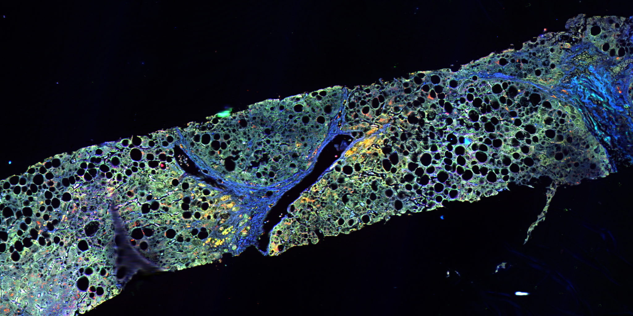
Virtual Stainer allows us to extract more information from each sample and to more richly annotate the intrinsic biology.
With no FDA-approved medications or treatments, NASH–a severe form of non-alcoholic fatty liver disease present in 3-12% of the adult U.S. population–can severely impact a person’s quality of life and deteriorate liver function to the point of requiring transplantation. In fact, NASH is predicted to be the primary cause of liver transplants by 2020. The current assessment of NASH disease activity is limited in the amount of information that is extracted from tissue biopsies, despite the inherently information-rich nature of histopathological samples. By obtaining a more quantitative and comprehensive picture of NASH biospecimens through Virtual Stainer, our collaboration hopes to discover signatures predicting disease progression and potentially new understanding of the disease and molecular pathways that, one day, could advance the treatment of this underserved condition.
“Today, liver biopsy is the gold standard used to diagnose NASH—the fastest growing liver disease worldwide—and to evaluate potential new treatments to reverse its course. Virtual Stainer gives us a unique opportunity to evaluate the richness of information available in liver tissue acquired through biopsy, well beyond currently available staining and imaging technologies,” says Dr. Laurent Fischer, Head of Liver Therapeutic Area at Allergan.
This collaboration to build our understanding of NASH is our first endeavor in applying Virtual Stainer to a specific disease area. At Verily, we believe that innovation in life sciences and healthcare is best achieved through the unification of diverse skills, and we are excited to see the work of our team of hardware engineers, optical physicists, software engineers, laboratory scientists, and clinicians come to fruition with Virtual Stainer. Starting with NASH, we are working to actualize the full potential of tissue collection to one day improve our knowledge of disease processes, with the goal of identifying better treatments for people on their journey back